Photodynamic therapy (PDT) has emerged as a promising modality for cancer treatment, but its efficacy is often limited by tumour hypoxia. Here, we report the development of a novel protein-based, self-assembled nanoplatform, CAT-I-BODIPY NPs (CIB NPs), to address this limitation. We first design and synthesize an I-BODIPY photosensitizer based on the heavy atom effect and modification of the electron-donating group, which exhibits excellent capabilities in generating reactive oxygen species and enabling near-infrared (NIR) fluorescence imaging. The incorporation of an oxygen-producing enzyme, catalase (CAT), within these nanoassemblies enables in situ oxygen generation to counteract hypoxic constraints. Controllable self-assembly by multiple supramolecular interactions into highly ordered architecture not only guarantees CAT's catalytic activity but also leads to excellent NIR fluorescence imaging ability and enhanced PDT efficacy. Notably, the visualization of optimal accumulation of CIB NPs within tumour sites 18 h post-injection offers precise PDT application guidance. Both in vitro and in vivo studies corroborate the remarkable anti-tumour efficacy of CIB NPs under NIR illumination, providing a significant advancement in PDT. The favourable biosafety profile of CIB NPs further emphasizes their potential for clinical application in hypoxic tumour therapy.
1. Introduction
The annual increase in cancer mortality has placed a tremendous burden on modern society, which necessitates the urgent development of innovative cancer therapies.1–3 Photodynamic therapy (PDT) has emerged as a promising cancer treatment modality with significant advantages, such as minimal invasiveness, low toxicity, negligible drug resistance, and excellent spatiotemporal selectivity.4–8 Among various photosensitizers (PSs) utilized for PDT, boron dipyrromethene (BODIPY) derivatives have attracted considerable attention due to their superior photochemical properties and potential as dual-function agents for both bioimaging and PDT.9–14 Particularly, the introduction of heavy atom iodine to BODIPY significantly enhances the photodynamic effect, mainly attributed to the heavy-atom effect, which improves spin–orbital coupling and facilitates the intersystem crossing rate, thus increasing the 1O2 QYs of PSs.15,16 Simultaneously, identifying the optimal treatment window is vital in phototherapy, as fluorescence imaging techniques allow for the visualization of the phototherapeutic process due to their high sensitivity, real-time monitoring capabilities, and noninvasive nature.17–20 Remarkably, iodinated BODIPY candidates also maintain satisfactory fluorescence imaging properties, making them suitable for imaging-guided PDT.16,21,22 Nevertheless, for these type I PSs, oxygen supply is essential to guarantee effective PDT, which may be restricted due to the intrinsically hypoxic tumour microenvironment.23–25 Furthermore, the inherent hydrophobicity of BODIPY easily leads to molecular aggregation, resulting in reduced therapeutic outcomes.26 To address these challenges and boost the selectivity and efficiency of PDT, there is an urgent need for the development of innovative PSs and strategies.27
The emergence of supramolecular self-assembly nanotechnology has provided an opportunity to propel the further development of efficient PDT.28–30 Through the orderly arrangement of various components, it can not only achieve multifunctional integration in one platform with increased stability but also allows for enhanced control over the physicochemical properties of the assembled nanostructures, thus improving therapeutic efficacy.31 To tackle tumour hypoxia, various strategies have been explored, including direct oxygen supply, enhancing intratumoral blood flow, and in situ oxygen production.32,33 Catalase (CAT), a natural enzyme with excellent biocompatibility, is an ideal choice for oxygen supply in tumours due to its ability to generate oxygen from hydrogen peroxide (H2O2).34–37 However, most studies focus on modifying CAT, which may compromise its efficiency and oxygen-generating ability.35,36 On the other hand, direct binding of functional moieties is more advantageous for improving the biosafety of the nanosystem compared to traditional encapsulation.38 Therefore, it is crucial to develop suitable strategies that effectively integrate proteins and PSs for safe and efficient tumor treatment.39,40
In this study, we developed a protein-based self-assembled nanoplatform (CAT-I-BODIPY NPs) for enhanced PDT of hypoxic tumours. The intriguing I-BODIPY PS, with remarkable ROS-generating ability and NIR-emissive fluorescence imaging capability, was synthesized by introducing heavy atom iodine and an electron-donating group 4-methoxybenzaldehyde. Oxygen-producing CAT with inherent biocompatibility was integrated into the nanoplatform to overcome hypoxic tumour microenvironments. Upon rational design and regulation of hydrophobic and hydrogen-bonding interactions, the self-assembly of CAT and I-BODIPY could be conducted to construct highly ordered nanoarchitectures. Under NIR laser irradiation, the real-time visualization by fluorescence imaging allowed for guiding the optimal time window for treatment. The ordered assembly of multi candidates well guaranteed the integration and functional maximization of each motif. Furthermore, the in situ oxygen generation of the nanoassemblies led to a significant promotion of the photodynamic effect and efficient cancer cell apoptosis. The innovative CAT-I-BODIPY nanoplatform addressed conventional PS limitations, providing a promising and biocompatible strategy for non-invasive hypoxic tumour treatment (Scheme 1).
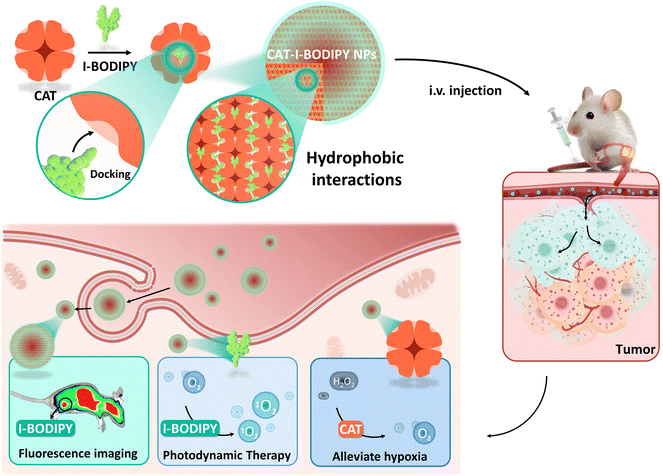
Scheme 1 The self-assembled nanomaterials of CAT and I-BODIPY with in situ oxygen-supplementation for enhanced PDT.
3. Results and discussion
3.1 Synthesis and characterization of B1–B4
To synthesize the I-BODIPY candidate (B4), we employed a four-step process involving a condensation reaction, two substitution reactions, and a Knoevenagel condensation reaction (Fig. 1a). Each of B1, B2, B3, and B4, when dissolved in DCM, presented distinct and vibrant colours under sunlight and UV light (Fig. S2†). The structures of B1, B2, B3, and B4 were confirmed through 1H NMR and mass spectrometry analyses (Fig. S3–S10†). After successful synthesis, we investigated the optical properties of these compounds. B1 and B2 showed similar absorption and fluorescence emission spectra, with λmax, Abs and λmax, Em both located at 500 nm and 545 nm, respectively (Fig. S11 and S12†). These similar spectra could be due to the unaltered position of the BODIPY core. B3 displayed a red-shifted emission peak and a roughly four-fold decrease in fluorescence intensity compared to B2 (Fig. S14†), possibly due to the incorporation of heavy atoms (iodine) at positions 2 and 6 of the BODIPY core. This modification may also boost the spin–orbit coupling effect in the molecule, thus enhancing the intersystem crossing rate and increasing the production of singlet oxygen (1O2) quantum yield (ΦΔ) by PSs. The addition of an electron-donating 4-methoxyphenyl substituent to the BODIPY core (B4) resulted in a redshift in the absorption and fluorescence emission spectra (Fig. S15 and S17†). The calculated ΦΔ was 73% and 67%, respectively according to the established methodology,41 indicating excellent PDT efficacy (Fig. 1b–e, Fig. S1†). Moreover, the redshift of B4 to the NIR region could effectively overcome the issue of tissue penetration depth, making it an ideal candidate for imaging-guided PDT.
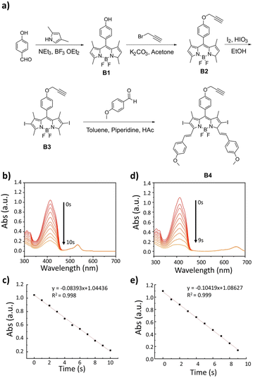
Fig. 1 (a) Synthetic routine for I-BODIPY. (b) The absorbance degradation of DPBF in the appearance of B3 as PSs under irradiation of Xe lamp. (c) Linear fitting of absorbance degradation in (b). (d) The absorbance degradation of DPBF in the appearance of B4 as PSs under irradiation of Xe lamp. (e) Linear fitting of absorbance degradation in (d).
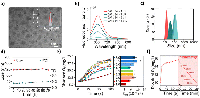
Fig. 2 Characterization of CIB NPs. (a) Transmission electron microscopy (TEM) image of CIB NPs at an equal concentration of CAT and B4 (CAT : B4 = 1 : 1) with a scale bar of 100 nm. The inset showed the dynamic light scattering (DLS) measurement of the CIB NPs at an equal concentration of CAT and B4 (CAT : B4 = 1 : 1). (b) Fluorescence spectra of CAT and B4 under different assembly conditions. (c) Assembly kinetics of CIB NPs. (d) Particle size and polydispersity index (PDI) of CIB NPs measured by DLS over 72 h. (e) Oxygen generation and catalytic efficiency of CIB NPs at different pH conditions and relative catalytic activity. (f) The dissolved oxygen content of CIB NPs after light irradiation.
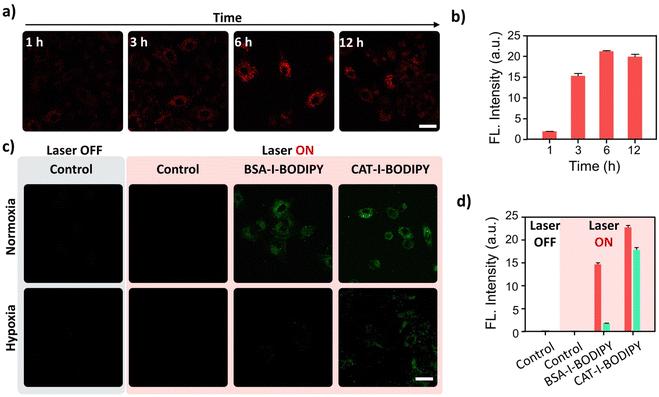
Fig. 3 (a) CLSM images of A549 cells were obtained after incubation with CIB NPs for different periods. (b) The relative fluorescence intensity of A549 cells in (a) was measured after incubation with CIB NPs at different times. (c) CLSM images of A549 cells were obtained after staining with SOSG following treatment with CIB NPs both in normoxic and hypoxic conditions, with or without 650 nm laser illumination. (d) The relative fluorescence intensity of A549 cells in (c) was measured after staining with SOSG following treatment with CIB NPs, with or without 650 nm laser illumination. Scale bar: 40 μm.
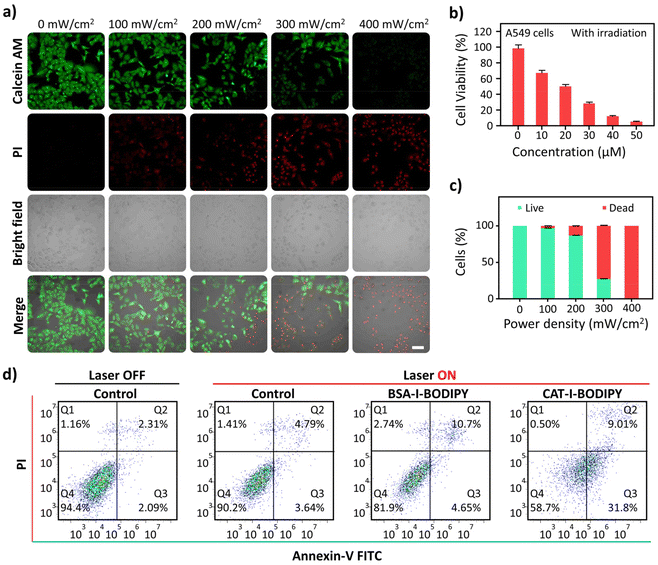
Fig. 4 (a) CLSM images of A549 cells treated with different light powers and stained with Calcein-AM and PI. Scale bar: 100 μm. (b) The effect of different concentrations of CIB NPs on A549 cell viability after 15 min of 650 nm light irradiation (300 mW cm−2) was tested by a CCK-8 assay. (c) The relative fluorescence intensity of A549 cells in (a) stained with Calcein-AM and PI after different treatments. (d) Flow cytometry assay of different treatments.
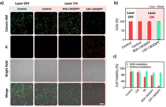
Fig. 5 (a) CLSM images of A549 cells stained with Calcein-AM and PI after conducting different treatments were displayed in hypoxic conditions. Scale bar: 100 μm. (b) The relative fluorescence intensity of A549 cells in (a) stained with Calcein-AM and PI after different treatments. (c) The effect of different groups (control, CAT, B4, BSA-I-BODIPY NPs, CAT-I-BODIPY NPS on A549 cell viability under hypoxic conditions after 15 min of 650 nm light irradiation (300 mW cm−2) was tested by a CCK-8 assay.
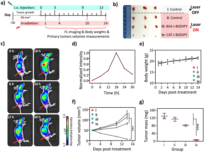
Fig. 6 In vivo application of CIB NPs. (a) Schematic diagram of the xenograft A549 tumour-bearing nude mouse model construction and in vivo NIR imaging-guided enhanced PDT plan in mice. (b) Images of tumours collected from different groups of nude mice 15 days after treatment. (c) Real-time fluorescence imaging was performed on A549 tumour-bearing nude mice after intravenous injection of CIB NPs. (d) The relative fluorescence intensity of in vivo fluorescence imaging in (c) was measured on A549 tumour-bearing nude mice after intravenous injection of CIB NPs. (e) The body weight of nude mice was measured after different treatments. (f) Tumor volume in nude mice was compared after various treatments. Data were presented as mean ± SD, ***p < 0.001. (g) The tumour mass of nude mice after different treatments is presented in mean ± SD (standard deviation), with statistical significance indicated as ****p < 0.0001.
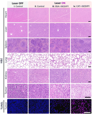
Fig. 7 H&E and TUNEL staining of tumour tissues and major organs from various treatment groups in nude mice. Scale bar: 100 μm.
4. Conclusions
In summary, this study presented a novel self-assembled bionanomaterial, CIB NPs, which offered a promising approach for imaging-guided enhanced PDT to hypoxic tumours. TEM and DLS well confirmed the successful construction of the nanoassemblies based on the hydrophilic protein and hydrophobic I-BODIPY. The assembled nanoprobe also retained its oxygen production capability, fluorescence imaging ability, and singlet oxygen generation capacity. Notably, it was observed that the nanoprobe reached maximum accumulation in the tumour site 18 h post-injection, providing a reliable basis for the implementation of precise PDT. In vitro and in vivo results on A549 demonstrated pronounced inhibition of tumour growth under NIR illumination due to the in situ supply of oxygen. Moreover, in vivo safety tests revealed favourable biocompatibility and negligible side effects in major organs, supporting the possible clinical application of CIB NPs in cancer treatment. This work will potentially pave the way for the development of more precise and effective diagnostic and therapeutic strategies in oncology, benefiting patients with hypoxic tumours.

