Fluorescent Chemosensors
Hardback ISBN:
978-1-83916-386-9
PDF ISBN:
978-1-83916-732-4
EPUB ISBN:
978-1-83916-733-1
Publication date:
14 Apr 2023
About this book
Fluorescent chemosensors have been widely applied in many diverse fields such as biology, physiology, pharmacology, and environmental sciences. The interdisciplinary nature of chemosensor research has continued to grow over the last 25 years to meet the increasing needs of monitoring our environment and health.
More recently, a large range of fluorescent chemosensors have been established for the detection of biologically and/or environmentally important species, and are increasingly being used to solve biological problems. The use of these molecules as imaging probes to diagnose and treat disease is gaining momentum with clear future applications.
This book will bring together world-leading experts to describe the current state of play in the field and introduce the cutting-edge research and possible future directions into fluorescent chemosensors design. Chapters focus on the basic principles involved in the design of chemosensors for specific analytes, problems, and challenges in the field.
Concentrating on advanced techniques and methods, the book will be of use for academics and researchers across a number of disciplines, with international appeal.
英国皇家化学会『超分子化学专著』系列丛书 (Monographs in Supramolecular Chemistry) 中的新书《荧光化学传感器》(Fluorescent Chemosensors) 已于 2023 年 4 月刚刚出版发行。
本书编辑为英国巴斯大学 Tony D. James 教授、吴庐陵博士、牛津大学 Adam C. Sedgwick 博士、华东理工大学贺晓鹏教授。本书汇集了全球专家学者的权威观点,有来自中国学者贡献了本书的第一、三、四、十一、十三章的内容。欢迎点击文末“阅读原文”查看本书网页。
荧光化学传感器已被广泛应用于诸如生物学、生理学、药理学和环境科学等许多领域。为了满足日益增长的监测环境和健康的需求,化学传感器研究的跨学科属性在最近25年间不断发展壮大。 目前,科学家已经创造了一系列荧光化学传感器,用于检测对生物学和环境重要的物种,并且越来越多地用于解决生物学问题。 使用这些分子作为成像探针来诊断和治疗疾病的势头越来越大,未来的应用也很明确。
本书汇集了全球专家学者的权威观点,总结荧光化学传感器设计领域的现状,介绍了最前沿研究成果并分析未来的方向。各章重点介绍针对特定分析物设计化学传感器所涉及的基本原理、该领域的问题和挑战。本书专注于先进的技术和方法,对于横跨多个领域的学者和研究人员大有裨益。
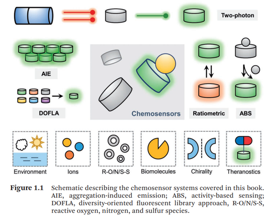
本书图 1.1: 本书涉及化学传感器体系的示意图
Luling Wu, (吴庐陵,英国巴斯大学) Adam C. Sedgwick, (牛津大学) Xiao-Peng He (贺晓鹏,华东理工大学) and Tony D. James* (英国巴斯大学)
Anthony W. Czarnik* (美国内华达大学雷诺分校)
- Chapter 3 活性传感:选择性生物成像的原理和探针
Shang Jia (贾上,美国加州大学伯克利分校) and Christopher J. Chang* (美国加州大学伯克利分校)
- Chapter 4 基于聚集诱导发光 (AIE) 的荧光体系
Meng Li,* (李檬,华北电力大学) Xiaoning Li and Zhijun Chen (陈志俊,东北林业大学)
- Chapter 5 面向多样性的荧光库方法:加速生物和环境应用的探针开发
Animesh Samanta,* (印度希夫纳达尔大学) Subrata Munan, Anal Jana and Young Tae Chang* (韩国浦项科技大学)
Vinayak Juvekar and Hwan Myung Kim* (韩国亚洲大学)
- Chapter 7 比率型荧光化学传感器:光物理/化学机理原理和设计策略
Jinwoo Shin, Jusung An, Jungryun Kim, Yuvin Noh, Paramesh Jangili* (高丽大学) and Jong Seung Kim* (高丽大学)
- Chapter 8 使用紫外-可见光吸收、荧光和圆二色光谱进行手性传感
James R. Howard, Jongdoo Lim, Sarah R. Moor and Eric V. Anslyn* (美国德克萨斯大学奥斯汀分校)
Michał Bartkowski and Silvia Giordani* (爱尔兰都柏林城市大学)
S. M. Butler and K. A. Jolliffe* (澳大利亚悉尼大学)
Ping Li* (李平,山东师范大学) and Bo Tang* (唐波,山东师范大学)
- Chapter 12 靶向亚细胞器的锌离子荧光探针
Toshiyuki Kowada and Shin Mizukami* (水上 進,日本东北大学)
- Chapter 13 用于硫烷硫和活性硒检测、成像的分子荧光探针
Zhenkai Wang, Feifei Yu, Yanlong Xing, Rui Wang, Heng Liu, Ziyi Cheng, Jianfeng Jin, Linlu Zhao* (赵琳璐,海南医学院) and Fabiao Yu* (于法标,海南医学院)
Tasuku Hirayama* (平山 祐,日本岐阜药科大学)
- Chapter 15 用于癌症治疗的可激型活光动力光敏剂
E. Kilic, M. Dirak and S. Kolemen* (土耳其科奇大学)
A. A. Bowyer and E. J. New* (澳大利亚悉尼大学)
- Chapter 17 延时镧系元素发光传感器和探针
Samuel J. Bradberry, Bruno D’Agostino, David F. Caffrey, Cidália M. G. dos Santos, Oxana Kotova and Thorfinnur Gunnlaugsson* (爱尔兰都柏林圣三一大学)
*本书出版时间为 2023 年 4 月,全书 490 页。如对本书感兴趣,请邮件至 RSCChina@rsc.org 咨询!随信附上编码:QJDW4RC1 可享受微信专属优惠!
- Chapter 13 用于硫烷硫和活性硒检测、成像的分子荧光探针
Zhenkai Wang, Feifei Yu, Yanlong Xing, Rui Wang, Heng Liu, Ziyi Cheng, Jianfeng Jin, Linlu Zhao* (赵琳璐,海南医学院) and Fabiao Yu* (于法标,海南医学院)
Abstract
Oxidative stress occurs when the body's oxidants and antioxidants are out of balance, which is considered to be one of the important factors leading to aging and disease. Antioxidants of non-enzymatic reactive chalcogenide species play an important role in redox homeostasis, among which sulfane sulfur species and active selenium species are particularly important. As a class of antioxidants with potential clinical biomarker value, the intracellular levels and distribution of sulfane sulfur and active selenium species can directly prove the dynamic state of oxidative stress, which may reveal the difference between physiological and pathological conditions. Fluorescence bioimaging technology has the advantages of high temporal and spatial resolution, low invasiveness and fast response, and has become a powerful tool for intracellular detection. Here, we summarize the design strategy and development of fluorescent probes for the detection of sulfane sulfur and active selenium in recent years.
Introduction
Since the concept of oxidative stress was added to the field of redox biology and medicine in 1985,1 the issues of oxidative stress are involving in physiology and pathophysiology research including chemistry, biochemistry, cell biology, as well as molecular biology in medicine field. Oxidative stress refers to the dysfunction between antioxidant system and reactive oxygen species (ROS) in organism. Under normal conditions, the intracellular milieu maintains redox homeostasis. ROS produced in a steady-state redox balance play important roles in biomolecules modification via oxidation-reduction (redox) reactions and then contribute to redox signaling. However, a chronic abnormal oxidative stress has been involved in diseases including cancer, Parkinson's disease, Alzheimer's disease, atherosclerosis, autoimmune diseases, inflammatory, reproductive system diseases, neurodegenerative diseases, and so on.2-4 Currently, the investigation on molecular redox switches manipulating oxidative stress responses has been elegantly established.5 Antioxidant defense is crucial for maintaining intracellular redox homeostasis. Although the major role in antioxidant defense is directly performed through antioxidant enzymes, the small molecular antioxidant compounds, as indispensable members, are involved in the elimination of reactive oxygen species via special manners. To prevent the organ systems against oxidative stress, the low molecular weight compounds such as ascorbic acid, vitamin A, vitamin E, carotenoids, coenzyme Q, α-tocopherol, uric acid, melatonin, aminoindoles, flavonoids, and polyphenols are behaving interactively and synergistically to remove of the excessive reactive oxygen species. In addition, another type of non-enzymatic antioxidants has attracted special attention in the field of redox homeostasis research, that is, reactive chalcogenide species, mainly containing reactive sulfur species and reactive selenium species, such as glutathione (GSH), cysteine (Cys), homocysteine (Hcy), hydrogen sulfide (H2S), α-lipoic acid, sulfite (SO32-), sulfane sulfurs, selenocysteine (Sec), hydrogen selenide (H2Se), and selenite (SeO32-). As a class of antioxidants with potential clinical biomarker value, the intracellular levels and distribution of reactive chalcogenide species can directly demonstrate the dynamic state of oxidative stress, which may reveal the different discrimination between physiological and pathological conditions. 6, 7
Since the reactive chalcogenide species is so vital to the health of living organisms, it is crucial to be able to evaluate the intracellular concentration accurately and sensitively. At present, the detection methods including high-performance liquid chromatography (HPLC), capillary electrophoresis, spectrophotometry, mass spectrometry (MS)/HPLC-MS have been elegantly established.8, 9 However, these detection methods require expensive instruments, invasiveness, tedious sample pretreatment procedures, and susceptibility to environmental interference. In particular, it cannot satisfy the in-situ and real-time inspection of analytes that are highly reactive, unstable in property and impossible to quickly separate from biological samples.
It is desirable that the visualization of the cells and organism accomplished with non-invasive technology, meanwhile, avoiding the body damage or disturbing of the distribution of intracellular constituents and isolating the intracellular analytes. Therefore, the techniques for imaging of level and distribution changes of biologically reactive species under the physiological or pathophysiological states become more than increasingly important in biomedical field. Since the fluorophores can be excited from a light source indwelling and their fluorescence emission detected without direct contact. Therefore, the whole measurement procedure can be operated non-invasively, in suit, and in real time. Fluorescence bioimaging technology based on fluorescent probes have been distinguished readily amongst biological detection technologies due to its several outstanding advantages in terms of high sensitivity, good selectivity, short response time, real-time monitoring, in situ observation, low cost, simple operation, high temporal and spatial resolution, and non-invasive measurement.8, 10-12 There have been many special summaries and discussions on biological reactive species, such as reactive oxygen species, reactive sulfur species, anions and cations, biological enzymes, and so on.13-15 As far as we know, there many entries of fluorescent probes related to the imaging and detection of sulfane sulfur and reactive selenium species have been reported. But there is few dedicated introductions on them. Herein, we summarize the current fluorescent probes for sulfane sulfur and reactive selenium species detection. These paradigms illustrate that fluorescent probe can serve as powerful tools for selectively detect sulfane sulfur and reactive selenium species in cells and in vivo, therefore suppling a promising approach for inspecting the physiological and pathological mechanisms. We expect to point out a path centered on fluorescence imaging for the detection of these high biological reactive species in living systems.
Conclusions and Perspective
The profound comprehensions of the signaling roles and physiological effects involved in redox homeostasis of sulfane sulfur and reactive selenium species are urgently required. The contributions of these species in generation and inhibition of redox processes by dominating biologically reactive species at nanomolar steady-state concentrations to levels which can trigger signaling pathway. The recognition of the preferred targets for sulfane sulfur and reactive selenium species and of their downstream signaling intermediates are not yet completely comprehended. The bio-functional validation of how these species affects individual proteins and regulate intracellular redox flux is still not available. Therefore, the lack of selectively, sensitive, accurate, and readily available chemical tools for quantifying sulfane sulfur or reactive selenium species proposes additional technical challenges.
The fields of redox biology benefit greatly by investigating the potential formations and decay kinetics and reactivities of sulfane sulfur or reactive selenium species in chemical properties. Appropriate chemical tools that are able to translate subtle variation into a non-invasive, in-situ, and in real-time visible signals are essential for diseases clinical diagnosis and therapy. Compared with other biological detection technologies, fluorescent biological imaging technology can provide researchers with a sensitive, real-time, high-precision testing results, which can also avoid the interference from intracellular endogenous species, so that giving the researchers an opportunity to monitor the level changes of reactive biological species more accurately for clinical diagnosis, therapy, and prevention. In this chapter, we summarize the current significant progress in the synthesis and design strategies of fluorescent probes used to detect sulfane sulfurs and reactive selenium species. The fluorescent probes used to detect sulfane sulfurs includes persulfide, polysulfide, hydrogen polysulfide. The fluorescent probes used to monitor reactive selenium species involves selenocysteine, hydrogen selenide, and thioredoxin. All these design strategies are derived from the nucleophilic addition reactions such as reduction, nucleophilic substitution, Michael addition, thiolysis, and so on.
Although the fluorescent probes currently for illustrating the vital roles of sulfane sulfurs and reactive selenium species in organisms behave excellent performance under physiological conditions, a few unsatisfied challenges always prevent such detection platforms from clinical applications. The fluorescence switch-on response makes the biological function inside the biological system more clearable, which intimately offers thorough exposition of these species functions with good spatial and temporal resolutions. This is one of the key issues for the clinical diagnosis. Analyte-specific probes can be beneficial to the accurate detection of the intrinsic behavioral process and trigger the discernable detection signals. Moreover, in order to guarantee the response signals emitting from the reaction site under native conditions, an effectively organelle targeting group must be included. The high specificity of molecular selective labeling must achieve the extended 3D imaging capability. It’s important to remember that conceiving a desirable fluorescent probe also requires that the ultimate fluorescence emission occurs within longer wavelength spectral range. Fluorophores with near infrared r excitation/emission wavelength can facilitate the deeper penetration, can minimize photodamage, and can avoid interference from intracellular autoluminescence. We should also clearly recognize that such fluorescent probe should address the existing limitations and realize to its potential clinical application.

